Статьи журнала - Гений ортопедии
Все статьи: 2037
Chondromas and multiple enchondromatosis
Статья научная
Introduction The chondromas are a cartilaginous proliferation of mature appearance and moderate size, reason why these tumors are regarded more like hamartomas than real benign tumor. Chondromas represent 10 to 12 % of benign bone tumors. Any bone of an enchondral ossification may be involved. Several bones can be involved, and the disease is called “chondromatosis”. In the review we describe clinical and radiological findings of this pathology as well as indications for reconstructive surgery. Material and methods The review is dedicated to isolated chondromas, periosteal and extraskeletal chondromas, chondromatosis. Results The aspects of epidemiology, clinical presentation, radiology, MRI, prognosis, indications and methods of surgical treatment have been described in the article for each types of chondroma and enchondromatosis. Conclusion Chondromas are benign bone tumors which may be responsible of pathologic fractures. Their surgical treatment consists in curettage and bone grafting or bone-cement filling with or without osteosynthesis. Multiple enchondromatosis should be considered as an osteochondrodysplasia. Its treatment is not the treatment of the multiple chondromas themselves, but of the bone deformities and length discrepancy induced by the disorder. The transformation of some tumors in chondrosarcomas in adolescence or adulthood needs a strict follow up of these patients.
Бесплатно
Статья научная
Background Chronic Chopart dislocation is one of the causes of acquired painful flat foot, which is treated by midtarsal arthrodesis causing limitation of movement and smaller-sized foot. Gradual reduction based on the principles of аrthrodiastasis using the Ilizarov external fixator is used for treating chronic Chopart dislocation. The case and method Twenty-two year-old male presented with painful right flat foot fourteen months after a motor vehicle accident. Gradual reduction was used for the chronically dislocated Chopart’s joint by arthrodiastasis using the Ilizarov external fixator. Result The follow-up result after four years is presented. The longitudinal arch of the foot recovered and the foot is painless with full range of movements; the size of the foot is preserved. Conclusion Treatment of chronic Chopart dislocation by arthrodiastasis using the Ilizarov external fixator is a preferred method of treatment as the size of the foot will be preserved , movement of the joint will not be restricted, the joint will be painless. There is no need for thromboprophylaxis, no chance of compartment syndrome and less operation time in comparison to arthrodesis.
Бесплатно
Статья научная
Introduction. Despite the excellent clinical success with total knee arthroplasty (TKA), there is disagreement about whether or not to replace the patellar articular surface. This led to randomized controlled trials. Such studies are the most reliable source of evidence of the effectiveness of potential interventions. But most of these studies include the study of results at all degrees of osteoarthritis of the patellofemoral joint. Therefore, the authors conducted a prospective study to compare the clinical and radiological outcomes after TKA with the replacement of the patellar articular surface in patients with osteoarthritis of the patellofemoral joint of the fourth degree. Materials and methods. The study included 123 patients with Kellgren-Lawrence patellofemoral joint osteoarthritis. Patients were randomly assigned to groups, in one of which the patellar joint surface was replaced (62 cases), in the other case, they were not performed, i.e. retained the patella (61 cases). Among them were 114 patients who were observed for more than two years (group with replacement of the articular surface - 59 cases, the group without replacement - 55 cases). Preoperative and postoperative clinical data were evaluated and evaluated on the scale of the Hospital for Special Surgery Patellar (HSSP) (total score of 100, presence of pain in the anterior part of the knee, functional limitations, tenderness in palpation or pressure, crepitation , Q-force). Scales developed at the Special Surgery Hospital (HSS), and the WOMAC scale, as well as the volume of movements (ROM), were also used. Results. The mean HSSP score in the group with replacement of the patellar articular surface was 85 points and 83 points in the group without replacement, which showed no significant differences between the groups (p = 0.75). When assessing the presence of pain in the anterior part of the knee, there were also no significant differences between the groups (40 in the group with articular replacement, 36 in the group without replacement, p = 0.52). HSS scores improved to 94 points in the group with articular replacement and up to 95 in the group without replacement, which also indicated no significant difference (p = 0.92). The WOMAC score and the volume of movements were 32 and 128 ° ± 10.5 ° in the replacement group and 29 points and 126 ° ± 11.5 ° - in the group without replacement, there was no significant difference between the groups (p> 05) . The conclusion. Thus, identical good clinical outcomes without significant differences were achieved after TKA with and without replacing the patellar articular surface in patients with a high degree of osteoarthritis of the patellofemoral joint. TKA without replacing the articular patella surface is a good option in patients with a high degree of osteoarthritis of the patellofemoral joint.
Бесплатно
Статья научная
Introduction To investigate clinical, laboratory findings and identify pre-operative variables associated with increased waiting time to surgery (WTS) and length of hospital stay (LOS) among hip fracture patients. Material and methods This prospective study is conducted between April 2020 and April 2021. Patients’ information was collected from medical records and subjected to analysis using a univariate and multivariate model. Results The study included 118 patients in a mean age of 79.5 years, and the majority were female (68.6 %). Overall, 66.9 % of the patients had at least one comorbidity. Almost all (95.8 %) patients had fractures due to a low-impact fall and an intertrochanteric fracture was the predominant type (61.9 %). The most abnormal laboratory findings at admission were elevated C-reactive protein (CRP) levels (94.9 %) followed by decreased mineral density (85.1 %), anaemia (81.4 %), electrolyte abnormalities (69.4 %) and hypoalbuminemia (66.1 %). The mean of WTS among 115 patients undergoing surgical treatment was 52.1 ± 47 hours and no patient-related factors had a significant influence on WTS. The mean hospital LOS was 15.9 ± 4.7 days. Marked elevation of CRP level (OR = 3.317, p = 0.042), type of surgery (OR = 4.413, p = 0.005) and WTS (OR = 4.602, p = 0.001) were independent predictors of prolonged LOS. Conclusion Most patients with hip fractures are elderly and suffer from many comorbidities and laboratory abnormalities. No patient-related factors are predictors of WTS but the elevation of CRP, type of surgery and time of waiting to surgery influence the LOS.
Бесплатно
Clubfoot: current concept of treatment
Статья научная
Introduction Idiopathic clubfoot (IC), also referred to as congenital talipes equinovarus, is one of the most common lower limb deformities observed in newborns, leading to significant functional impairment when left untreated. Early minimally invasive treatment has been praised as one of the most successful practice of modern pediatric orthopedics. This review aims to report current knowledge and controversies about clubfoot treatment. Material and methods We describe the main trends in clubfoot managing, identifying peculiarities, difficulties and prognostic factors related to the treatment. Results Many treatment techniques either conservative, surgical or hybrid have been used over the past decades. Based on good and excellent results during long-term follow-up, Ponseti method has been globally accepted by paediatric orthopaedic surgeons as standard method of treatment. However, some other conservative methods are still widely applied in the clinical setting, such as the French Physical Therapy method. Adherence to the bracing protocol is critical for the long-term success of the treatment, being a better predictor for relapse than severity of the deformity at birth. Conclusions Taking care of the manipulation and casting details by trained professionals, together with enhancing the child and patents’ adherence to the brace, are essential for the success of conservative treatment. Surgery should be performed only when strictly needed, preferably on a “a la carte” approach.
Бесплатно
Статья научная
Background Fixation of pathological long bones with telescopic intramedullary rods is well known to be a technically challenging procedure even in specialist centres, with a high complication rate due to rod migration, hardware failure, nonunion or malunion. However there is very little guidance in the literature regarding salvage treatment options when failure occurs.Aim We demonstrate a surgical technique that can be used for salvage treatment of both femoral and humeral complex nonunions following Fassier-Duval (FD) rodding in a child with osteogenesis imperfecta (OI).Case description A 13 year-old girl with OI type VIII presented sequentially with nonunion and deformity of the femur then the humerus following previous FD rods in those segments. The femur was also complicated with metallosis between the steel rod and an overlying titanium plate. Both segments were treated with pseudarthrosis debridement, removal of metalwork and stabilisation with hydroxyapatite (HA)-coated flexible intramedullary nails, with temporary Ilizarov frame to provide enough longitudinal and rotational stability to allow immediate weight-bearing. The femur Ilizarov frame was removed after 64 days, and the femur remained straight and fully healed at 2.5 years. The frame time for the humerus was 40 days, complete union was achieved and upper limb function restored and maintained at 9 months.Discussion The transphyseal telescopic rod is the traditional implant of choice in terms of treating fractures and stabilising osteotomies for deformity in OI. However, it does not provide enough torsional or longitudinal stability by itself to allow early weight-bearing which is detrimental to bone healing in this vulnerable patient group. The incidence of delayed union or nonunion at osteotomy site in telescopic rod application is not negligible: up to 14.5-51.5 %. Although the technique we have shown in this case may not be applied to all complex OI patients, we believe that the combination of flexible intramedullary nails and Ilizarov frame provides a favourable environment for bone healing in complex or revision cases. As a secondary learning point the initial revision surgery to the left femur demonstrated the perils of using a steel rod and a titanium plate in a biologically active environment which in this case lead to metallosis and lysis.Conclusion We found the technique of HA-coated flexible intramedullary nails combined with the Ilizarov frame effective in the salvage of failed telescopic rods in both femur and humerus and feel this technique can be used as a salvage option in similar cases worldwide. This case also demonstrates the perils of using different metals in combined internal fixation.
Бесплатно
Статья научная
Introduction Spinal tuberculosis is an extra-pulmonary tuberculosis infection caused by Mycobacterium tuberculosis (MTB) which affects the vertebrae. Culture test is the «gold standard» diagnostic method, but it takes a long time. PCR is a method of a shorter time than the culture test, so it can be an alternative diagnostic method for MTB. Materials and Methods This study is a cross-sectional study. The data were analyzed with diagnostic tests on patients with suspected spinal tuberculosis who performed surgery in Hasan Sadikin Hospital in Bandung. Clinical examinations and diagnostic examinations were done in 40 patients and surgery was performed to obtain samples from the spinal cord and the infected tissue. GeneXpert, PCR targeting IS6110 and culture tests were performed. The research was conducted at the Department of Orthopaedics and Traumatology and the Clinical Pathology Laboratory of FK UNPAD/RSHS from September 2019 to September 2020. Results GeneXpert assay compared with culture tests as the standard diagnostic method showed sensitivity of 96.67 %; specificity of 90.00 %; positive predictive value 96.67 %; and a negative predictive value of 90.00 %, with an accuracy of 95.00 %. The PCR targeting IS6110 against culture showed that the sensitivity of MTB bacterial infection was 93.33 %, the specificity was 80.00 %, the positive predictive value was 93.33 %, the negative predictive value was 80.00 %, and the accuracy was 90.00 %. Conclusion This study concluded that the GeneXpert MTB/RIF RT-PCR assay has a high sensitivity, specificity, and accuracy compared to PCR targeting IS6110 in examining tissue samples in patients with spinal TB.
Бесплатно
Статья научная
Introduction Osteoporosis is a disease causing high morbidity with increasing prevalence. It is one of chronic diseases caused by reduced bone mass that subsequently decreases bone strength and increases fracture risks. Pharmacologic treatments for osteoporosis include antiresorptive agent (bisphosphonate) and bone-forming agent (strontium ranelate), so further research is needed to compare these two medications. Objectives We aimed to histopathologically compare bone density in post-menopause white rats after being treated with strontium and ibandronate. Material and methods 45 ovariectomized female rats were divided into three groups. The subjects in the first group were only ovariectomized (control). The strontium group was given daily oral strontium at a dose of 625 mg/kg BW/day for 60 days. The ibandronate group was given one subcutaneous ibandronate injection at a dose of 1 μg/kg BW/day for 60 days. We measured osteoclasts, osteoblasts, trabecula area and cortical thickness. Results The animals in ibandronate and strontium groups showed a significant increase.
Бесплатно
Статья научная
Background Volar locking plate (VLP) has gained the most popularity in the treatment of distal radius fractures due to its superior biomechanical property. In contrast, external fixation (EF) is not so extensively used. The aim of this study was to find what procedure is better in the management and achieves favorable outcomes in patients with comminuted distal radial fractures. Patient and methods This study included 30 patients with distal radial fractures AO types A3, C2, C3 in which 15 ubjects were managed with open reduction and internal fixation by volar plate, and another 15 were managed with external fixation augmented by K-wires. The minimum duration of follow-up in our study was six months. Results Patients treated with external fixation augmented by K-wires had grip strength range 15-27, patients treated with volar plating had grip strength range 8-27. There was no significant statistical difference between 2 groups regarding extension. In Group A Mann-Whitney test revealed that Gartland-Werely score had negative correlation with affected hand. In Group B the correlation was positive with AO/OTA classification only and negative with affected hand and ulnar styloid fracture but also not statistically significant between Quick-DASH score with affected hand, AO/OTA classification and ulnar styloid fracture. Conclusion Both volar plating and external fixation augmented by K-wires are treatment choices for distal radius fractures. Whereas external fixation maintains a significant role in the treatment of distal radius fractures, ORIF with locked volar plating has changed the way many surgeons treat certain types of distal radius fractures.
Бесплатно
Статья научная
Introduction Re-infection rates remain high after revision for periprosthtic joint infection (PJI). We conducted a complex diagnostic study of hip PJI and studied bone tissue involvement into the infectious process. Material and methods Twenty-nine patients treated for PJI were examined. Ten patients had acute, seven had late and 12 hematogenous infection (Tsukayama, 1996). Clinical, laboratory, microbiological, radiographic and histological methods were used for PJI detection. Results ESR exceeded the threshold in 16 and CRP in 23 cases. Pathogenic microorganisms were confirmed in 23 cases. Radiographic manifestations of periprosthetic bone destruction were seen in 14 patients with late and hematogenous infection. The histological study showed signs of osteomyelitis in 19 patients, including five cases with acute PJI. Histological study of periprosthetic and pseudosynovial membranes revealed PJI in 20 patients. The histological and microbiological tests together confirmed PJI in 27 cases (92 %). ROC analysis showed that the accuracy of CRP in the diagnosis of osteomyelitis exceeded that of ESR due to its higher sensitivity. Microbiological tests showed satisfactory sensitivity but low specificity. The radiographic study had an extremely low sensitivity and low specificity. Histology of membranes was quite sensitive and specific for the diagnosis of osteomyelitis but did not reach the level of bone histology. Conclusions Our complex diagnostic study has enabled to accurately characterize the septic process in revision arthroplasty. The histological findings show that osteomyelitis of different severity might develop in each PJI type. This fact may be a key factor for a surgeon in choosing a more reliable treatment option.
Бесплатно
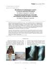
Congenital pseudoarthrosis of the tibia and treatment using the Ilizarov technique
Статья научная
Background: Congenital pseudoarthrosis of the tibia (CPT) is one of the most difficult problems in paediatric orthopaedic surgery. This is a rare condition, which is mostly associated with neurofibromatosis. Treatment options have varied greatly. Successful surgical treatment of tibial pseudoarthrosis is possible by the Ilizarov method. Patients and methods: 7 patients with CPT treated using the Ilizarov technique within the period from 1998 to 2008. RESULTS: Bone healing was achieved in all cases except one. (Union rate - 85,7 %). Conclsion: The Ilizarov technique is a comprehensive approach to all CPT aspects. Its use can end with very good results
Бесплатно
Статья научная
Emergency procedures aimed at rapid reduction and fixation and spanning of periarticular fractures has been termed “damage control orthopaedics”. In severely injured patients, early definitive fixation of fractures may not be appropriate. Recent studies showed that in multiple trauma, DCO is the best option for management of patients who are unstable and in extremis. The paper presents a case of such a control in a 34-year-old patient who sustained polytrauma on 20.10.2016. Primary medical care was conducted at a local hospital. Ten hours after the injury, the patient was transported to the Bari-Ilizarov orthopaedic centre for further management. On admission, he was in a traumatic shock. Radiographic study showed a comminuted fracture of the left femur, medial condylar fracture of the ipsilateral femur, comminuted fractures of both bones of the shin, left wrist sprain, and contusion of the head. Osteosynthesis of the left femoral shaft was performed with a Kuntscher nail and additionally with the Ilizarov fixator. When patient’s condition stabilized on the next day, osteosynthesis of the tibia was performed with the Ilizarov apparatus and the wrist was fixed with a plaster cast.
Бесплатно
Статья научная
Introduction Distal humerus fractures are relatively rare and being intra-articular are difficult to manage. As the number of elderly people grows steadily and also with increasing use of motor vehicles in the developing countries, it can be said that the frequency of intraarticular fractures of the distal humerus will increase similar to the fractures of the distal end of the radius, hip, and spine. There are several treatment plans for managing intraarticular fractures of the distal humerus depending on fracture anatomy. We conducted this study to assess which approach is superior, closed percutaneous reduction with K-wires or ORIF. Methods A total of 30 patients who satisfied the inclusion criteria were included, out of which 16 patients underwent ORIF and 14 patients underwent closed reduction and percutaneous pinning (СRPP). Patients included were between 21-50 years of age with intraarticular nonpathological closed distal humerus fractures without preoperative neurovascular deficit and presented less than 10 days between the fracture event and treatment. Results In our study, mean age of patients undergoing CRPP was 28.1 years while the mean age of patients undergoing ORIF was 30.1 years. This study showed that distal humerus fractures are more common in younger age groups. In our study, mean arc of motion at 6 months postoperatively in patients which underwent CRPP was 106.07 degrees while the mean arc of motion in patients which underwent ORIF was 80.94 degrees. In patients who underwent ORIF, only 6.2 % (1/16) had excellent outcome, 56.3 % (9/16) patients had good outcomes, 31.3 % (5/16) patients had fair outcomes, and 6.2 % (1/16) had poor outcome. It was found that out of a total of 14 patients which underwent CRPP, only 7.1 % (1/14) had cutaneous impingement. Fracture union occurred in 100 % of patients except 3 patients in ORIF group; however, they had partial union up till 6 months of follow-up. Conclusion Our study concludes that even displaced intra-articular fractures of the distal humerus can be satisfactorily treated with closed reductions and percutaneous pinning.
Бесплатно
Статья научная
Distraction osteogenesis according to Ilizarov is the most common method of bone lengthening and deformity correction including the hand and foot bones. This method forms the basis of the main techniques used for the hand bone lengthening. The author described the technical specifications of the mini-fixator developed for short tubular bones, demonstrated the diagrams of its application to finger phalanges and metacarpal bones, as well as he presented the illustrative clinical cases. The hand bone lengthening performed in 394 patients (566 segments of the hand) at the Center within the period of 1999 and 2012. The performed lengthening procedure improved the hand appearance and functions.
Бесплатно
Drainage versus non-drainage in primary total knee replacement: a comparative study
Статья научная
Goal. Compare the results in 80 patients who underwent total knee replacement, find out the advantages of using closed suction drainage and compare the results with those in the group where drainage was not used. Methods. A randomized prospective study was conducted in 2011 in group A (40 patients) without drainage and in group B (40 patients) with drainage. Total blood loss was significantly greater in patients who were drained (400 ml) compared to 200 ml in the group without drainage, although the latent amount of blood loss can not be accurately determined. Results. The statistical difference in terms of postoperative pain, ecchymosis or the frequency of cases of infection in the postoperative period in both groups was not. Conclusions. There were no clear evidence to support the use of drainage with total arthroplasty of the knee, despite this many surgeons prefer to use them.
Бесплатно
Experience of arthroscopic surgery in tophaceous gout: indications, results and complications
Статья научная
Background Gout, lasting 5 years or more, and high uncontrollable levels of uric acid in blood lead to the formation of tophi - gouty stones containing the UA crystals surrounded with connective tissue. As the result of tophi formation in the joint area patients felt extreme discomfort and quite often completely lose ability to work. Objectives To define indications for tophaceous gout surgery in the Chengdu Rheumatism Hospital, evaluate surgical results and complications, as well as the effectiveness of a new surgery equipment. Materials and methods The indications and results of tophaceous gout surgery were investigated in 63 male gout patients of Chengdu Rheumatism hospital in 2019-2020. A retrospective analysis was carried out on the basis of medical records for all patients who were prescribed with urate lowering therapy and underwent arthroscopic intervention or complex surgical intervention combining arthroscopic shaving with open tophectomy procedure. Results The most common lesion site was foot joints: toes (49.41 %), ankle (39.68 %) and knee (34.92 %), with restricted mobility in the mentioned joints. Among common complaints were inability to perform daily routines due to enlarged joints (inability to wear shoes), joints’ dysfunction and pain. Younger patients (aged 20-44) had significantly higher levels of uric acid in serum before treatment. In most cases, indications for surgery for this group of patients were pain and discomfort in joints, inability to perform daily work. After accessing pain levels, 38.46 % of younger patients reported pain leveled 6 or higher on VAS score, which was more often, compared to patients aged 45-55 (26.92 %) and older than 55 (10.0 %). After surgery and following urate lowering therapy all patients noted functional improvement and reduction of pain. Decrease in serum urate levels were reported in 96.83 % of patients. Conclusion The results of surgical treatment for functional impairment of the joint (inability to perform daily work due to restricted range of motions) and massive joint transformation (inability to wear shoes/clothes) in gout patients are positive, with all patients reporting functional improvement and reduction of pain, and the risk of complications is low. In addition to urate lowering therapy we cautiously recommend performing arthroscopic shaving even in younger gout patients consistent with aforementioned indications.
Бесплатно
Experimental and clinical studies on the reduction of bone lengthening terms at the Ilizarov center
Статья обзорная
Introduction The list of pathological conditions that need surgical correction of bone length is very long. In the literature on the topic under discussion, it is reported that the best results are obtained by lengthening according to the Ilizarov method. But many surgeons are not satisfied with the long duration of external fixation, which requires a large number of adjustments and patient compliance. The aim of this work is a analysis of the modern technologies for limb lengthening that actually shorten the time of osteosynthesis using the apparatus for external fixation. Materials and methods Literary sources have been analyzed since the first publication on limb lengthening according to Ilizarov in 1963. The search was carried out in the databases of the RSCI, NCBI Pubmed, Medline. The developments of the employees of the Ilizarov Center are presented. Results An analysis of the literature showed that the fixation units of the apparatus, units for providing movement for distraction, compression and correction of angles have been improved. The invasiveness of the surgical intervention is minimized. Best results can be obtained using automatic lengthening. The Ilizarov Center has developed and experimentally proven methods for reducing the period of distraction and the period of fixation with the apparatus. We combined three factors: an increased round-the-clock distraction rate (2 or 3 mm) with a motorized distractor adjusted to the Ilizarov frame and fixation reinforcement along with regeneration stimulation with HA-coated intramedullary wires in our experimental and clinical trials. This technology has easily conquered a number of clinical practices in Russia, France and Serbia. Conclusion Experimental and clinical substantiation of a two- or three-fold increase in the rate of automated distraction in the conditions of intramedullary reinforcement with a bioactive implant can drastically reduce not only the time of the distraction period, but also the duration of the fixation period with the Ilizarov apparatus.
Бесплатно
Femur head necrosis as a post-acute sequela of COVID-19 (Sars-Cov-2 infection)
Статья научная
Introduction While the COVID-19 pandemic may still be ongoing, we have simultaneously entered into the post-acute phase of COVID-19, which comes with its own challenges. This case series reports 11 patients of COVID-19 treated with corticosteroids who subsequently developed osteonecrosis of the femoral head (ONFH). Methods All consecutive patients diagnosed on MRI with ONFH from August 2020 to May 2021 and were retrospectively COVID-19 positive were included. The treatment administered for COVID-19 was retrieved and evaluated. The patients were managed for femoral head necrosis, and results were reported. Results Overall, 11 patients developed ONFH in a total of 16 hips. The severity of femoral head necrosis depended on the dose of corticosteroid administered during COVID-19. A high dose for a longer duration resulted in a higher ONFH stage (FICAT & Arlet ). Hips in the lower grade were treated conservatively, and in the higher grade were treated surgically. The follow-up scores of patients demonstrated steady improvement. Conclusions High suspicion of femoral head necrosis has to be considered in patients treated with corticosteroids for COVID-19 as it can aid in early detection and early intervention to preserve the native femoral head.
Бесплатно
Fibroblast growth factor receptor 1 extracellular vesicle as novel therapy for osteoarthritis
Статья научная
Introduction Osteoarthritis (OA) is a joint condition that causes significant impairment of the chondrocyte. The gradual degradation of the cartilage lining of one or more freely moving joints, as well as persistent inflammation, are the causes of osteoarthritis. Current medication focuses on alleviating symptoms rather than curing the condition. Methods This review article was completed by searching for information with the keywords “Fibroblast Growth Factor Receptor-1”, “Extracellular Vesicle”, and “Osteoarthritis” in various journals in several search engines. Out of 102 publications found, 95 were suitable to be studied. Results The upregulated amount of fibroblast growth factor receptors (FGFR1) signaling suggesting the progression of degenerative cartilage that commonly seen in osteoarthritis (OA) patients. Several studies showed that the involvement of extracellular vesicles (EV) derived from MSCs could enhance cartilage repair and protect the cartilage from degradation. EVs have the potential to deliver effects to specific cell types through ligand-receptor interactions and several pathway mechanisms related with the FGFR1. EVs and FGFR1 have been postulated in recent years as possible therapeutic targets in human articular cartilage. Conclusions The protective benefits on both chondrocytes and synoviocytes in OA patients can be achieved by administering the MSC-EVs that may also stimulate chondrocyte proliferation and migration. EVs have a promising potential to become a novel therapy for treating patients with OA. However, further research is needed to discover possible application of this therapy.
Бесплатно

